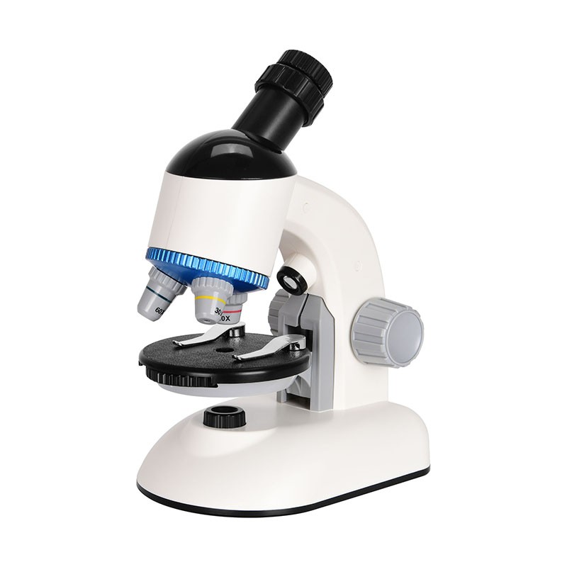
When using natural light source for microscopic examination, it is best to use a north-facing light source, not direct sunlight; When using artificial light sources, it is advisable to use the light source of fluorescent lamps.
During the microscopic examination, the body should face the practice table, adopt a correct posture, open the eyes naturally, observe the specimen with the left eye, observe the recording and drawing with the right eye, and adjust the focus with the left hand to make the object clear and move the field of view of the specimen. Right-handed recording, drawing.
The stage should not be tilted during microscopic examination, because when the stage is tilted, liquid or oil can easily flow out, which not only damages the specimen, but also contaminates the stage, and affects the inspection results.
During microscopic examination, the field of view of the specimen should be moved in a certain direction until the entire specimen is observed, so that the examination is not missed and not repeated.
The heavy light of the microscope is the conversion of the light, the objective lens and the adjustment of the light. Light conditioning is important when observing parasite specimens. Because the observed specimens such as worm eggs, cysts, etc., are natural light state objects, large and small, dark and light color, some colorless and transparent, and low magnification, high magnification objective lens conversion more, so it is necessary to adjust the focus and light at any time with different specimens and requirements during microscopic examination, so that the observed object can be clear. In general, the light of stained specimens should be strong, and the light of colorless or unstained specimens should be weak; The light observed by the low magnification mirror should be weak, and the light observed by the high magnification mirror should be strong.
1. To light:
(1) Turn the low-magnification lens to the bottom of the lens barrel and form a straight line with the lens barrel.
(2) Toggle the reflector to adjust to the brightest field of view without shadows. The reflector has two sides, flat and concave, flat when the light source is strong, concave surface when dark, and when strong light is needed, the concentrator is raised and the aperture is enlarged; When low light is required, lower the concentrator or reduce the aperture appropriately.
(3) Place the specimen to be observed on the stage, and turn the coarse adjuster to lower the lens barrel to the objective lens close to the specimen. While turning the coarse adjuster, lean over the mirror to carefully observe the distance between the objective lens and the specimen.
(4) The left eye is observed in the eyepiece, and at the same time, the left hand rotates the rough adjustment, so that the lens barrel slowly rises to adjust the focal length, so that the object in the field of view stops when it is seen, and then adjusts the micro-adjuster until the specimen is clear.
2. Use of objective lens and light adjustment:
Microscopes generally have three objective lenses, namely low-magnification, high-magnification and oil lenses, fixed in the nosepiece changer hole. When observing the specimen, use a low-magnification objective lens first, at this time, the field of view is larger, the specimen is easier to detect, but the magnification is small (generally 100 times), and the structure of the smaller object is not easy to observe. High-magnification objective lenses have a large magnification (typically 400x magnification) and can observe tiny objects or structures.
The worm eggs of parasites, microfilariae, trophozoites and cysts of protozoa, and larvae of insects all use low and high magnification. Protozoa in tissue cells, oil mirrors are used. Use low and high magnification to observe, if the object or its internal structure cannot be accurately identified under low magnification, turn to high magnification lens observation. Using an oil lens to observe, generally add a drop of oil and directly immerse the oil lens into the oil droplet for microscopic observation.
3. Recognition of low-magnification, high-magnification and oil lenses:
(1) Indicate the magnification of 10×, 40×, 100 ×, or 10/0.25, 40/0.65, 100/1.25.
(2) The low magnification lens is the shortest, the high magnification lens is longer, and the oil lens is the longest.
(3) The mirror hole in front of the lens has the largest low magnification lens, the high magnification lens is larger, and the oil lens is the smallest.
(4) The oil lens is often engraved with a black ring, or the word "oil".

4. How to use low magnification lens for high magnification lens:
(1) After the light is right, move the thruster to look for the specimen that needs to be observed.
(2) If the size of the specimen is large and its structure cannot be clearly detected and therefore cannot be confirmed, move the specimen to the center of the field of view, and then rotate the high-magnification objective lens under the lens barrel.
(3) Rotate the micro-regulator until the object is clear.
(4) Adjust the concentrator and aperture to make the objects in the field of view reach the clearest degree.
5. How to use the oil mirror:
(1) Principle: When using an oil mirror to observe, you need to add cedar oil, because the oil mirror needs to enter the lens with more light, but the gas permeability of the oil mirror is the smallest, so that the light entering is less, and the object is not easy to see clearly. At the same time, due to the light transmitted from the slide, refractive astigmatism occurs due to the density of the medium (slide-air-objective lens) (slide: n=1.52, air: n=1.0), so less light enters the lens and the object is more unclear. Therefore, a medium similar to the refractive index of the slide, such as cedar oil, is used between the specimen and the slide, so that the light does not pass through the air, so that more light enters the lens and the object can be seen clearly.
(2) The use of oil mirror:
a. Turn the light to its maximum intensity (the concentrator is raised, the aperture is all open).
b. Turn the coarse adjuster to raise the lens barrel and drop 1 small drop of cedar oil (not too much, do not spread) on the specimen just below the objective lens.
c. Turn the nosepiece adapter so that the oil lens is under the lens barrel.
d. Under the observation of the naked eye, turn the coarse adjuster to slowly lower the oil lens and immerse itself in the cedar oil, until it gently touches the slide.
e. Slowly turn the coarse adjuster so that the oil lens slowly rises until the object of the specimen is seen.
f. Turn the micro-adjuster to make the visual field of view the clearest degree.
G. Slowly move the thruster with the left hand and turn the micro-adjuster to observe the specimen.
h. After the specimen is observed, turn the coarse adjuster to raise the lens barrel and remove the specimen slide. Immediately wipe the citronella oil off the lens with lens paper.
6. Precautions:
(1) Before using the microscope, you should be familiar with the names and methods of use of each part of the microscope, especially the characteristics of identifying three types of objective lenses.
(2) Most of the specimens observed in parasitology practice are colorless and light-colored, so attention must be paid to the adjustment of light.
(3) When observing fresh specimens, a coverslip must be added to prevent the specimen from drying and deforming due to evaporation or pollution to erode the objective lens, and at the same time make the surface of the specimen uniform, and the light can be concentrated, which is conducive to observation.


